-
Products
-
Transportation & Mobility Solutions
Transportation & Mobility Solutions
At Hitachi, we engineer industry-leading transportation and mobility solutions by leveraging decades of knowledge and using high-quality automotive material and components.
-
Energy Solutions
Energy Solutions
We believe the only solution for fulfilling the growing power requirements of industries and society is through a comprehensive portfolio of sustainable energy solutions and delivering innovative high-efficiency energy systems.
-
IT Infrastructure Services
IT Infrastructure Services
Hitachi’s state-of-the-art IT products and services are known to streamline business processes which result in better productivity and a higher return on investment (ROI).
- Application Modernization
- Consulting Services
- Data Modernization
- Data Storage
- Data-Driven Industrial Operations
- DataOps
- Digital Transformation
- Edge to Cloud Infrastructure Services
- Industrial Data & IoT Management
- Industry Solutions
- Infrastructure Management & Analytics
- Infrastructure Modernization
- Managed Security
- X-as-Service - Everflex
-
Social Infrastructure: Industrial Products
Social Infrastructure: Industrial Products
Within the industrial sector, Hitachi is consistently delivering superior components and services, including industrial and automation solutions, useful in manufacturing facilities.
-
Healthcare & Lifesciences
Healthcare & Lifesciences
At Hitachi, we believe that healthcare innovation is crucial to a society’s advancement. A strong healthcare sector is often considered an inseparable element of a developed society.
-
Scientific Research & Laboratory Equipments
Scientific Research & Laboratory Equipments
Hitachi focuses on extensive research and development, transformative technology, and systems innovation to unfold new possibilities and create new value through scientific endeavors that strengthen the connection between science and social progress.
-
Smart Audio Visual Products
Smart Audio Visual Products
Since 1956, Hitachi audio visual products have provided state of the art solutions to consumers all over the world. It has been our pleasure to design competitive products at the lowest possible prices while maintaining our industry-leading quality standards for your comfort and enjoyment.
-
View All Products
Hitachi Products & Solutions
Hitachi, a technology leader in the U.S., offers a diverse set of products and solutions, and breakthrough technologies for smart manufacturing, green energy and mobility solutions that empower governments, businesses, and communities.
-
Transportation & Mobility Solutions
- Social Innovation Solutions
-
About Us
-
Hitachi in the U.S.A.
Hitachi in the U.S.A.
Discover information about the Hitachi group network across the Americas, upcoming events and sustainability endeavours, CSR policies, and corporate government relations.
-
About Hitachi Group
About Hitachi Group
Explore our leadership team, investor relations, environmental vision, and sustainability goals. Learn how Hitachi is leveraging its research & development capabilities for social innovation across industry verticals.
-
Hitachi in the U.S.A.
- News Releases
- Case Studies
- Careers
- R&D
FLOW VISUALIZATION TAKES CENTER STAGE AT TEDMED 2016 AS DR. PARTHO SENGUPTA, IN COLLABORATION WITH HITACHI, PRESENTS ADVANCED VISUALIZATION TECHNIQUES
Wallingford, CT, December 5, 2016 - Evaluation of cardiac hemodynamics progresses to a new level as presented by Dr. Partho Sengupta at TedMed 2016. In collaboration with Hitachi and others, Dr. Sengupta demonstrated a true ground-breaking technology; the evaluation of cardiac valves using holograms.
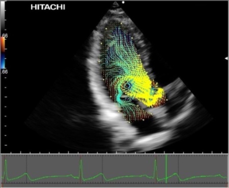
His presentation tells his personal story of an impressive journey through the diagnostic world of cardiac imaging that ultimately brought him to where he is today. His focus is strongly on discovering advanced visualization technologies to evaluate valvular form and function and how it is linked to the cardiac flow geometry. Some of Dr. Sengupta’s past work has been with PIV to demonstrate the valvular flow with vector information. He shared how he is now also using ultrasound to provide the vector information to evaluate this same flow geometry. He presents examples of PIV and Hitachi’s Vector Flow Mapping (VFM) analysis results side by side to illustrate the validity of ultrasound as a tool for vector mapping.

VFM using Velocity Vectors and Streamline display in a normal patient
As a look into the future, Dr. Sengupta then demonstrated how holograms will play a part in valvular and other cardiac diagnosis by showing an example of a mitral valve hologram. This example provided a remarkable snapshot into the future of evaluating cardiac physiological characteristics by using holograms.
Hitachi’s VFM stands in the forefront as an example of advanced hemodynamic analysis today. In the future, VFM along with hologram analysis are true leading-edge technologies that will certainly advance the diagnostics of cardiac health.


